Urinary Stones
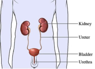
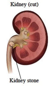
Urine contains many minerals and salts. If these elements become concentrated and reach critical levels, they precipitate in your urine to form stones. After forming in the kidney, the stones may migrate down into other parts of the urinary tract including the ureter, bladder, and urethra. As they pass through the ureter, they may cause pain and blood in the urine. Passing a stone is described as one of the most painful events a human may experience in his / her lifetime! If the stone blocks the flow of urine, the kidney is at risk of developing permanent damage. In addition, life threatening infections may develop.
Stones vary in shape and size. When they remain small, they can be excreted without pain. We may place you on medication to assist the passage of stones, or to try to dissolve them. In other cases, surgery may be needed. Treatment at our Kidney Stone Center, a state-of-the-art facility, is focused on removing kidney stones and preventing existing stones from growing. We also emphasize lifestyle changes to prevent new stones from forming. We provide complete blood and urine evaluation to identify the cause of stone formation and are aggressive in helping you prevent stones in the future.
We are experts in the evaluation and treatment of kidney stones. We offer only the safest, most advanced stone treatment options. Almost all stones are treated endoscopically (surgery on the inside of the body, without the need for cutting the patient) or by other Minimally Invasive Surgeries. We feature ureteroscopic and Laser approaches, ShockWave Lithotripsy, and Percutaneous Nephrolithotomy (the safest, most sophisticated treatment available for large kidney stones).
Treatment Options
-
Extra-Corporeal Shock Wave Lithotripsy
-
Ureteroscopy (URS)
-
Percutaneous Nephrolithotomy (PCNL)
-
Prevention of Stones
<
>
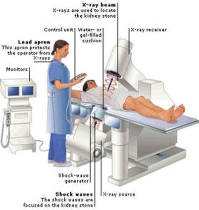
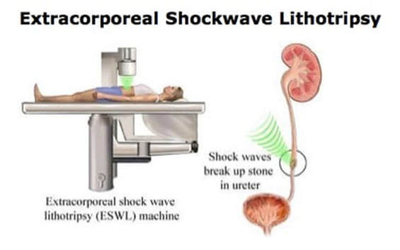
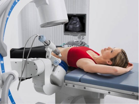
Shock Wave Lithotripsy (ESWL) is used to treat stones in the kidney and ureter. Shock waves are generated by an external machine, which uses X-rays to locate the stone. The shock waves are focused on the stone, and with repeated firing the stone eventually breaks into small pieces. These small pieces will pass out in your urine over the next few weeks, painlessly.
Because of possible mild to moderate discomfort caused by the shock waves and the need to control your breathing during the procedure, some form of anesthesia is often used. This may range from local anesthesia to sedation or general anesthesia.
SWL does not work well on hard stones, very large stones, or stones that do not show up on X-ray (radiolucent stones). If you are pregnant, ESWL cannot be performed on you because of the risk to your foetus.
With ESWL, you go home on the same day as the procedure. We offer a mobile EWSL unit that comes to the hospital of your preference island-wide to treat you. You should be able to resume normal activities in two to three days.
Although SWL is widely used and considered very safe, it can still cause side effects. You may have blood in your
urine for a few days after treatment. Larger stone fragments may get stuck in the ureter, causing pain and needing other removal procedures.
Click here to see information video for ESWL
Although SWL is widely used and considered very safe, it can still cause side effects. You may have blood in your
urine for a few days after treatment. Larger stone fragments may get stuck in the ureter, causing pain and needing other removal procedures.
Click here to see information video for ESWL
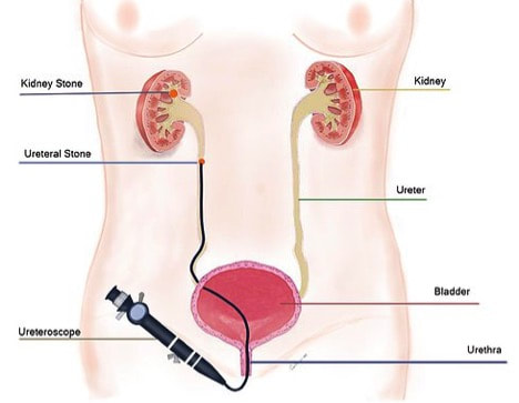
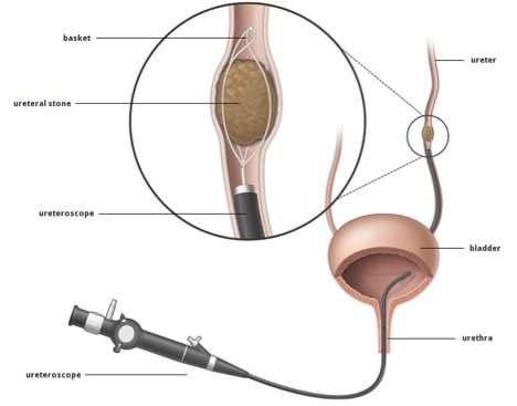
Ureteroscopy (URS) is used to treat stones in the kidney and ureter. URS involves passing a very small telescope, called an ureteroscope, into the bladder, up the ureter and as far as the kidney if necessary. Rigid telescopes are used for stones in the lower part of the ureter near the bladder. Flexible telescopes are used to treat stones in the upper ureter and kidney.
The ureteroscope lets us pass a camera inside your body via your natural orifice (urethra), without having to make an incision (cut). We can then operate on the stone totally internally. General anesthesia is required so that you feel no pain during the procedure. Once the stone is visualized, a small, basket-like device grabs smaller stones and removes them. Larger stones are broken into smaller pieces with a laser or other stone-breaking tools.
Once the stone has been removed whole or in pieces, we may place a temporary stent in the ureter. A stent is a tiny, rigid plastic tube that helps hold theureter open so that urine and stones can drain from the kidney into the bladder. This tube is completely internal (within the body). It does not require an external bag to collect urine. It does not affect your daily activites, and you can return to work the next day.
You usually are able to go home the same day as the URS. If a stent was placed, it is removed about one week later. This is a painless procedure that is performed in the office without the need for anesthesia.
Percutaneous Nephrolithotomy (PCNL) is the safest, most efficient treatment for large stones in the kidney. General anesthesia is necessary to perform a PCNL.
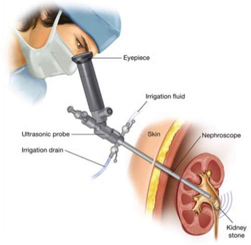
PCNL involves making a 1 cm incision (cut) in the side, just large enough to allow a rigid telescope (nephroscope) to be passed into the part of the kidney where the stone is located.
PCNL involves making a 1 cm incision (cut) in the side, just large enough to allow a rigid telescope (nephroscope) to be passed into the part of the kidney where the stone is located.
An instrument passed through the nephroscope breaks up the stone and suctions out the pieces.
After the PCNL, a tube (nephrostomy) may be left in the kidney to drain urine into a bag outside the body. It is not our practice to routinely leave any form of tube or stent after PCNL, in order to improve your after-surgery comfort level and decrease complication rates. If a tube is necessary, it is left in overnight or for a few days.
You usually have to stay in the hospital overnight after this operation. You can begin normal activities after discharge home and may return to work shortly after. Strenuous physical activities/ sports will have to be postponed for a few weeks, however.
Prevention of Stones
After your stones have been treated, we actively focus on maintaining your stone- free status. Stone –formers are at a 50% lifetime risk of forming stones again in the future. We perform stone, urine and blood analyses to learn what type of stones you have formed, to better educate and advise you on life-style and dietary changes necessary to prevent any future events.

